Your body creates about 25 million new cells every second. Most of these cells form through mitosis, a precisely choreographed process that ensures each new cell receives an exact copy of your genetic material. Understanding what happens during mitosis is fundamental to grasping how organisms grow, heal wounds, and replace worn-out tissues. This guide breaks down each stage of mitosis so you can master the process for your next exam or lab report.
Mitosis divides one cell into two genetically identical daughter cells through five distinct phases: prophase, prometaphase, metaphase, anaphase, and telophase. The process ensures accurate [DNA replication](https://www.ncbi.nlm.nih.gov/books/NBK9876/) and distribution, allowing multicellular organisms to grow and repair tissues. Following mitosis, cytokinesis physically separates the two new cells. Understanding each phase helps you recognize how cells maintain genetic stability across generations.
Why Cells Need Mitosis
Cells cannot grow indefinitely. As a cell increases in size, its surface area to volume ratio decreases, making it harder to transport nutrients in and waste out efficiently. Mitosis solves this problem by creating two smaller cells from one large cell.
Multicellular organisms rely on mitosis for three main purposes. Growth happens when your body produces more cells than it loses. A baby grows into an adult because trillions of mitotic divisions create new cells for bones, muscles, organs, and skin. Repair occurs when damaged tissues need replacement cells. When you scrape your knee, mitosis generates new skin cells to close the wound. Replacement maintains tissues that naturally wear out, like the lining of your digestive tract, which completely renews every few days.
Single-celled organisms use mitosis differently. For them, mitosis equals reproduction. When an amoeba undergoes mitosis, it creates two separate organisms.
The Cell Cycle Context
Mitosis represents just one phase of the complete cell cycle. Before a cell can divide, it must prepare properly.
The cell cycle consists of two major periods: interphase and the mitotic phase. Interphase takes up about 90% of the cycle and includes three stages. During G1 phase, the cell grows and performs its normal functions. S phase is when DNA replication occurs, doubling the genetic material. G2 phase allows the cell to continue growing and prepare the machinery needed for division.
Only after completing interphase does a cell enter the mitotic phase, which includes mitosis itself and cytokinesis. This careful sequencing prevents cells from attempting division before they have copied their DNA.
What Happens During Prophase
Prophase marks the beginning of mitosis and involves dramatic changes to cell structure. The replicated DNA, which spent interphase as loosely packed chromatin, now condenses into visible chromosomes. Each chromosome consists of two identical sister chromatids joined at a region called the centromere.
The nucleolus disappears during prophase. This structure, normally visible inside the nucleus, fades as the cell redirects its resources toward division.
Outside the nucleus, the centrosomes move to opposite poles of the cell. These organelles serve as organizing centers for the mitotic spindle, a structure made of microtubules that will eventually separate the chromosomes. As the centrosomes migrate, microtubules begin extending from them, forming the early spindle apparatus.
The nuclear envelope starts to break down late in prophase, transitioning into the next stage.
Prometaphase Prepares for Alignment
Some textbooks treat prometaphase as part of prophase, while others describe it separately. Either way, this stage involves critical preparations.
The nuclear envelope completely fragments into small vesicles. With the barrier gone, spindle microtubules can now access the chromosomes directly.
Each chromatid develops a protein structure called a kinetochore at its centromere. Kinetochores serve as attachment points for spindle microtubules. Some microtubules successfully attach to kinetochores, while others extend from one pole to the other or interact with microtubules from the opposite pole.
Chromosomes begin moving, pulled by the attached microtubules. This movement appears chaotic at first, with chromosomes shifting back and forth as the cell establishes proper attachments.
Metaphase Achieves Perfect Alignment
Metaphase is often the easiest stage to identify under a microscope because of its distinctive arrangement. All chromosomes align at the cell’s equator, forming what scientists call the metaphase plate.
This alignment is not random. Each chromosome is connected to microtubules from both poles, creating tension that pulls the sister chromatids in opposite directions. The chromatids remain joined at the centromere, so they stay in place at the cell’s center.
The cell uses a checkpoint mechanism during metaphase to verify proper attachment. If any chromosome lacks correct microtubule connections, the cell pauses mitosis until the problem is fixed. This checkpoint prevents errors that could result in daughter cells receiving the wrong number of chromosomes.
Anaphase Separates Sister Chromatids
Anaphase begins suddenly when the proteins holding sister chromatids together break down. The centromere splits, and the former sister chromatids, now individual chromosomes, move toward opposite poles.
Two types of movement occur simultaneously. Kinetochore microtubules shorten, pulling chromosomes toward the poles. At the same time, the poles themselves move farther apart as the cell elongates.
Anaphase is the shortest stage of mitosis, typically lasting only a few minutes. By its end, two identical sets of chromosomes occupy opposite ends of the cell.
Telophase Reverses Early Changes
Telophase essentially reverses the events of prophase. The chromosomes arrive at the poles and begin to decondense, returning to their extended chromatin form.
Nuclear envelopes reform around each set of chromosomes, creating two separate nuclei. The nucleolus reappears in each new nucleus.
The spindle apparatus disassembles as the cell no longer needs it. Microtubules break down, and their components will be recycled for other cellular uses.
By the end of telophase, the cell contains two nuclei but still exists as a single cell. The final separation requires cytokinesis.
Cytokinesis Completes Cell Division
Cytokinesis overlaps with telophase but is technically separate from mitosis. This process physically divides the cytoplasm and organelles between the two daughter cells.
Animal cells accomplish cytokinesis through a contractile ring. Actin and myosin filaments form a ring around the cell’s equator and contract, pinching the cell membrane inward. The indentation deepens until the cell splits into two, similar to pulling a drawstring tight.
Plant cells cannot pinch inward because their rigid cell walls prevent it. Instead, they build a new wall from the inside out. Vesicles carrying cell wall materials move to the cell’s center and fuse, forming a structure called the cell plate. The plate expands outward until it connects with the existing cell wall, dividing the parent cell into two daughter cells.
Each daughter cell receives approximately half the cytoplasm and organelles. While the genetic material is divided exactly, organelle distribution is more random. Cells typically contain enough organelles that random distribution works fine.
Key Differences Between Mitosis and Meiosis
Students often confuse mitosis with meiosis, but these processes serve different purposes and produce different results. Understanding the distinctions helps clarify what happens during mitosis specifically.
| Feature | Mitosis | Meiosis |
|---|---|---|
| Purpose | Growth, repair, asexual reproduction | Sexual reproduction |
| Number of divisions | One | Two |
| Daughter cells produced | Two | Four |
| Chromosome number in daughters | Same as parent (diploid) | Half of parent (haploid) |
| Genetic variation | Identical to parent | Different from parent |
| Where it occurs | Somatic (body) cells | Germ cells (sex cells) |
Mitosis maintains chromosome number. If a human cell with 46 chromosomes undergoes mitosis, both daughter cells will have 46 chromosomes. Meiosis, by contrast, reduces the chromosome number by half, which is necessary for creating sex cells.
Common Student Mistakes When Learning Mitosis
Recognizing typical errors helps you avoid them in your own studying and test taking.
Many students confuse the order of phases. The sequence is prophase, prometaphase, metaphase, anaphase, telophase. Creating a mnemonic device helps. Some people use “Please Poke My Arm Today” or make up their own memorable phrase.
Another mistake involves mixing up chromatids and chromosomes. Before S phase, each chromosome consists of a single DNA molecule. After replication, each chromosome consists of two sister chromatids joined at the centromere. During anaphase, the chromatids separate and are then called individual chromosomes.
Students sometimes forget that mitosis refers only to nuclear division, not the complete process of creating two cells. Cytokinesis must follow mitosis for cell division to finish.
Diagrams can mislead if you do not pay attention to detail. A cell in metaphase shows chromosomes at the center, but this does not mean the DNA is still replicating. Replication finished during S phase, long before mitosis began.
Regulation and Control of Mitosis
Cells do not undergo mitosis randomly. Complex regulatory mechanisms control when and how often cells divide.
Cyclins and cyclin-dependent kinases (CDKs) act as the primary control system. Cyclin levels rise and fall throughout the cell cycle, and when they bind to CDKs, they trigger specific events. For example, high levels of certain cyclins push the cell from G2 phase into mitosis.
Checkpoints serve as quality control stations. The G1 checkpoint verifies that the cell is large enough and has adequate nutrients. The G2 checkpoint confirms that DNA replication completed successfully and that DNA is undamaged. The metaphase checkpoint, also called the spindle checkpoint, ensures all chromosomes are properly attached to spindle microtubules before allowing anaphase to proceed.
Growth factors from outside the cell also influence division rates. When you cut yourself, platelets release growth factors that signal nearby skin cells to divide more frequently, speeding wound healing.
When Mitosis Goes Wrong
Errors in mitosis can have serious consequences. If chromosomes do not separate properly during anaphase, one daughter cell receives too many chromosomes while the other receives too few. This condition, called aneuploidy, usually kills the cell or prevents it from functioning normally.
Cancer represents mitosis gone terribly wrong. Cancer cells ignore normal regulatory signals and divide uncontrollably. They bypass checkpoints that would normally stop division if problems are detected. Understanding normal mitosis helps researchers develop treatments that target the abnormal division patterns in cancer cells.
Some chemicals and radiation damage the mitotic spindle. Colchicine, for example, prevents microtubule formation, which stops cells at metaphase because they cannot separate their chromosomes. While this sounds harmful, doctors actually use spindle-disrupting drugs to treat certain cancers, since rapidly dividing cancer cells are more vulnerable to these agents than most normal cells.
Practical Study Strategies for Mastering Mitosis
Memorizing the phases is just the start. You need to recognize each stage visually and understand what is happening at the molecular level.
Create your own diagrams rather than just studying existing ones. Drawing forces you to recall details and understand spatial relationships. Label the chromosomes, spindle fibers, centrosomes, and cell membrane in each phase.
Use microscope slides or online image databases to practice identifying phases in real cells. Textbook diagrams are simplified. Actual cells look messier, and learning to recognize phases in authentic images prepares you for lab practicals.
Watch time-lapse videos of cells undergoing mitosis. Seeing the process in motion reinforces how one phase flows into the next. Many universities and educational organizations provide free microscopy videos online.
Study with a partner and quiz each other. One person describes a phase without naming it, and the other identifies which phase is being described. This technique builds both your recall and your ability to explain concepts, which helps during essay exams.
Connecting Mitosis to Other Biological Concepts
Mitosis does not exist in isolation. It connects to many other topics you will encounter in biology courses.
DNA replication, which you might study in a separate unit, is essential for mitosis. Without accurate replication during S phase, mitosis cannot produce genetically identical daughter cells. If you understand why do atoms form bonds, you can better appreciate the chemical interactions that hold DNA strands together and allow enzymes to copy them.
Cell signaling pathways regulate when cells enter mitosis. Growth factors bind to receptors on the cell surface, triggering cascades of molecular events inside the cell. These pathways involve many of the same principles you learn when studying how electronegativity determines molecule behavior and bond types.
Energy metabolism supports mitosis. Dividing cells need large amounts of ATP to build new structures and power molecular motors like the ones that move chromosomes. The principles behind what happens during an exothermic reaction apply to the energy-releasing reactions that fuel mitosis.
“Understanding mitosis is not about memorizing isolated facts. It is about seeing how DNA replication, protein synthesis, energy metabolism, and structural changes work together in a coordinated process. When you can explain why each step must happen in a specific order, you have truly mastered the material.”
Laboratory Observations of Mitosis
Seeing mitosis under a microscope transforms it from an abstract concept to a visible reality. Most biology courses include a lab where you observe mitotic stages in prepared slides.
Onion root tips are popular specimens because their cells divide frequently. The growing root constantly produces new cells, so a thin slice of root tip contains cells in various mitotic stages. After staining to make chromosomes visible, you can scan the slide to find examples of each phase.
Whitefish blastula slides show mitosis in animal cells. The blastula is an early embryonic stage with rapid cell division, providing many mitotic figures in a small area.
When examining slides, spend time on each cell. Prophase cells show condensed chromosomes but still have a visible nuclear envelope (early prophase) or fragments of it (late prophase). Metaphase cells display the characteristic line of chromosomes across the center. Anaphase cells show V-shaped chromosomes moving apart. Telophase cells have chromosomes clustered at opposite ends and often show a pinching in the middle from cytokinesis.
Count how many cells you find in each stage. Stages that take longer will be represented by more cells. You will probably find many cells in prophase and interphase but fewer in anaphase because it proceeds so rapidly.
Mitosis in Different Organisms
While the fundamental process remains similar across species, some variations exist.
Fungi undergo closed mitosis, where the nuclear envelope stays intact throughout division. The spindle forms inside the nucleus, and chromosomes separate within this enclosed space. Only after mitosis completes does the nucleus divide into two.
Most animal and plant cells perform open mitosis, with the nuclear envelope breaking down as described earlier. This allows the spindle to form in the cytoplasm and interact directly with chromosomes.
Some protists show intermediate forms, with the nuclear envelope partially breaking down or developing holes that allow spindle microtubules to enter.
These variations remind us that evolution has found multiple solutions to the same problem of dividing genetic material accurately.
The Role of Mitosis in Medicine and Research
Understanding mitosis has practical applications beyond passing your biology exam.
Doctors use mitotic index (the percentage of cells undergoing mitosis) as a diagnostic tool. Tumors with high mitotic indices tend to be more aggressive because the cancer cells are dividing rapidly. This information helps determine treatment approaches.
Prenatal testing sometimes involves examining cells from amniotic fluid. Technicians culture these cells and then stop them at metaphase, when chromosomes are most condensed and visible. This allows geneticists to count chromosomes and check for abnormalities.
Agricultural scientists study mitosis to develop better crops. Understanding how plant cells divide helps researchers create varieties that grow faster or produce more cells in edible parts like fruits.
Regenerative medicine depends on controlled cell division. Scientists working on growing organs or tissues must precisely regulate mitosis to produce the right number of cells in the correct arrangements.
Tools That Help Visualize Mitosis
Modern technology has given us powerful ways to observe and study mitosis.
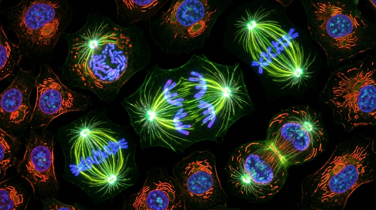
Fluorescent tags allow researchers to label specific proteins with glowing markers. By tagging tubulin (the protein that makes up microtubules), scientists can watch spindle formation in living cells using fluorescence microscopy. Different colored tags on different structures let them track multiple components simultaneously.
Time-lapse microscopy captures images at regular intervals, then plays them back at higher speed. This turns a process that takes an hour into a video lasting a minute, making it easier to see the flow from one phase to the next.
Computer simulations model the forces acting on chromosomes during mitosis. These models help researchers understand how cells achieve the precise movements needed for accurate chromosome separation.
Even simple light microscopy remains valuable. The same type of microscope used in introductory biology labs has allowed scientists to observe mitosis for over a century, and it still serves as an excellent learning tool.
Building Your Mitosis Knowledge Base
Start with the big picture, then add details. Understand that mitosis divides the nucleus and produces two identical daughter cells. Then learn the five phases in order. Finally, fill in the specific events within each phase.
Make connections between what you are learning and real-world examples. When you heal from a cut, that is mitosis. When a plant grows taller, that is mitosis. When your hair gets longer, that is mitosis. These connections make the material more memorable and meaningful.
Test yourself regularly rather than cramming before exams. Spacing out your study sessions helps move information from short-term to long-term memory. Try explaining mitosis to a friend or family member who has not studied biology. If you can make it clear to someone else, you truly understand it.
Pay attention to the language used in questions. “What happens during metaphase?” requires a different answer than “What is the purpose of metaphase?” or “How can you identify metaphase in a microscope image?” Practice answering questions phrased in different ways.
Why Mastering Mitosis Matters for Your Biology Education
Mitosis appears repeatedly throughout biology courses. In genetics, you learn how mitosis ensures offspring cells receive complete genetic information. In development, you study how mitosis contributes to an organism growing from a single fertilized egg. In evolution, you examine how errors in mitosis can introduce variation. In medicine, you explore how cancer represents uncontrolled mitosis.
Every time you encounter these topics, your understanding of mitosis provides the foundation. Students who thoroughly learn mitosis early find later topics easier because they already understand this fundamental process.
The skills you develop while learning mitosis also transfer to other subjects. Breaking a complex process into distinct phases, understanding cause and effect relationships, and integrating multiple levels of organization (molecular, cellular, organismal) are skills you will use throughout your scientific education and career.
Your investment in truly understanding what happens during mitosis pays dividends far beyond a single test grade. Take the time to master each phase, understand why each step must occur in sequence, and recognize how the process connects to other biological phenomena. Your future self, facing more advanced biology courses, will thank you for building this solid foundation now.